The underlying cause of a venous leg ulcer (VLU) is venous disease. Not everyone with vein problems will go on to have a leg ulcer, but everyone with a venous leg ulcer will have signs and symptoms of venous disease that they can trace back over time2.
Epidemiology
Venous leg ulcers are a significant public health concern in Australia and New Zealand, particularly among older adults. The overall prevalence of VLUs in the Australian population is approximately 1%, rising to 4% among individuals aged over 65 years3. In New Zealand, similar demographic patterns are observed, with VLUs predominantly affecting older adults and those with chronic venous insufficiency. VLUs are associated with high recurrence rates—up to 56% within three months of healing3 and prolonged healing times, with 50% remaining unhealed for over nine months4.
The lifetime risk of developing a venous ulcer is estimated at 10–20 per 1000 people, and the condition imposes a substantial economic burden, with annual treatment costs exceeding $1 billion in Australia5. Risk factors include age, immobility, obesity, and a history of deep vein thrombosis. The burden of VLUs is compounded by their impact on quality of life, frequent recurrence, and the need for long-term management strategies.
Aetiology
Venous leg ulcers are primarily caused by chronic venous insufficiency (CVI), which results from impaired blood flow in the veins of the lower leg, leading to venous hypertension. This condition arises when the one-way valves within the veins, responsible for preventing backflow of blood, fail to function properly, causing blood to pool in the lower leg and increasing pressure within the veins. This elevated pressure can then force fluid and blood cells out of the veins and into the surrounding tissues, damaging the skin and eventually leading to ulcer formation6.
The pathology can also include venous obstruction (e.g. from blood clotting).6 Venous drainage is impaired, which will lead to venous hypertension7,8. Between 40–50% of venous leg ulcers are due to superficial venous insufficiency combined with perforating vein incompetence, but the deep vein system4 is usually normal.
Diagnosis of chronic venous insufficiency is based on clinical characteristics; chronic venous hypertension causes a number of skin changes:
- oedema or swelling
- visible capillaries around the ankle
- trophic skin changes which are abnormalities of the skin from insufficient nerve or blood supply including hyperpigmentation (discolouration) or hemosiderin staining (dark brown discolouration
- atrophie blanche (white discolouration with reddish dots)
- Induration (injury) of the skin and underlying tissue9 lipodermatosclerosis)
- stasis eczeme (venous eczema, dry scaly inflamed skin)9
In patients with chronic venous insufficiency, the inability of the calf muscles to pump venous blood contributes to the development and delayed healing of venous leg ulcers. As a result, compression treatment is used to treat lower limb venous insufficiency5,10.
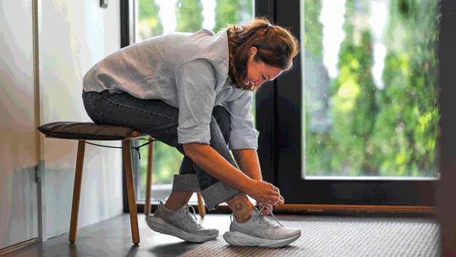
There are many risk factors for venous leg ulceration, including heredity, obesity, venous occlusion, and age4,10. Importantly, venous leg ulceration reoccurs in up to 70% of people who are at risk4. More than 95% of venous leg ulceration is in the leg below the knee, usually around the malleoli, and ulceration may be discrete or circumferential11.
Clinical and economic burden
Venous leg ulcers significantly impact Australia's economy, with an estimated annual cost of $3 billion12. This includes both direct healthcare costs and out-of-pocket expenses for individuals13.
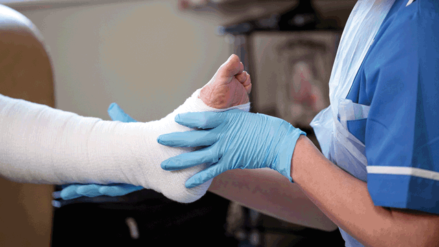
There has been prior advice to admit patients to hospital for continuous leg elevation to improve healing14 and clinical judgement may deem this appropriate. However, recent strategies of management in the community is common. Management in the community, as home or clinic-based care is more cost-effective, reduces reliance on hospital services, and is often preferred by patients, provided there are no major complications, significant comorbidities, or impaired mobility preventing access to community services. While specialized hospital care may be necessary for unhealed ulcers, severe cases, or patients requiring intensive intervention, a collaborative approach involving trained community nurses is often more sustainable and improves access to VLU services15.
Too often, early preventive treatment is not undertaken due to the increasing number of patients with venous leg ulcers and the increasing shortage of hospital beds, the high cost of in-patient hospital care, and the need to maintain independence in the mainly elderly population who suffer from venous leg ulcers15.
Effects on patient quality of life
Venous leg ulceration is often a chronic condition, and patients experience a prolonged cycle of skin healing and then breakdown (read more about the challenges of this cycle). This cycle of wound healing and reversal sometimes repeats over decades, with episodes of infection, all of which can impair quality of life16.
Optimal management and care of venous leg ulcers: Psychosocial impacts and pain responses
Management of venous leg ulcers occurs mainly in the community, but community nurses and general practitioners have limited time to spend with patients, and this time is usually directed to clinical management16. Psychological and social effects of venous leg ulceration have received little attention in clinical management guidelines but include social isolation, anxiety, and depression, particularly when ulcers are highly exudative and painful16.
Optimal management and care of venous leg ulcers: Dressing selection and the M.O.I.S.T. model
As with most wound types, venous leg ulcers benefit from appropriate dressing selection and implementing a M.O.I.S.T. healing model.
Selecting dressings and M.O.I.S.T. management complement each other. Topical wound therapy options, such as dressings, are selected based on the goals and actions for healing, such as creating a balanced moist wound healing environment or improving tissue oxygenation, which are two of the key properties of the M.O.I.S.T. model.
The M.O.I.S.T. model is an acronym that stands for:
- Moisture balance: Creating a balanced moist wound healing environment
- Oxygen balance: Improving tissue perfusion and local oxygenation at the wound bed
- Infection control: Avoiding wound infection or treating an existing infection
- Supporting strategies: Creating a supportive wound environment to stimulate healing
- Tissue management: Removing devitalised tissue and debris, to form healthy granulation and epithelial tissue
M.O.I.S.T. principles can be applied across a range of chronic (hard-to-heal) wounds, including VLUs, and the application of M.O.I.S.T. has been shown to help such wounds “progress toward healing or achieved complete healing”17.